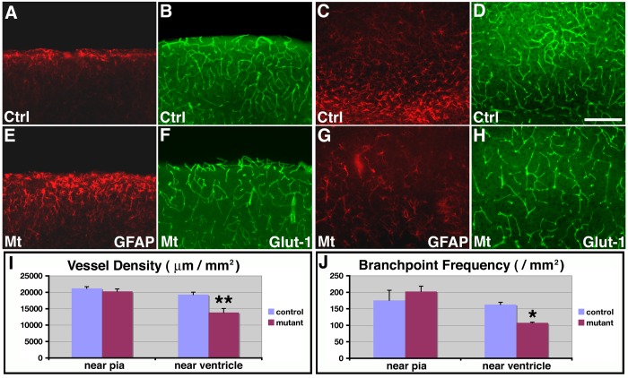
Figure 8. Recovery of vessel development in the upper cortex of class II orc3/hGFAP-cre mutants at later postnatal stages.
(A–H) Astrocytes and glial progenitors were stained using anti-GFAP antibodies (red in A, C, E & G) and blood vessel morphology was assessed by anti-Glut-1 staining (green in B, D, F & H) in control (A–D) and class II mutant (E–H) cortices at P14. Dramatic increases in GFAP expression in astrocytes near the pia were observed in mutants as compared to controls (compare E to A), while the number of glial progenitors near the ventricle remained relatively depleted in mutants (compare G to C). On the other hand, while vessel density remained low near the ventricle in mutants (compare H to D), vessel density in the upper cortex has recovered to normal in mutants (compare E to B). (I–J) Quantitative analysis of vessel density (I) and branching frequency (J) in control and mutants at P14. In cortical regions near the ventricle, significantly lower vessel density (**) (control, 16.7±30.4/mm2; mutant, 13837±1216 μm/mm2; P = 0.008, n = 4) and branching frequency (*) (control, 162.7±7.0/mm2; mutant, 107.5±1.9/mm2; P = 0.011, n = 4) were observed in mutants as compared to controls. By contrast, in regions near the pia, both vessel density (control, 21189±506 μm/mm2; mutant, 20271±736 μm/mm2; P = 0.35, n = 4) and branching frequency (control, 175.7±30.4/mm2; mutant, 202.3±16.2/mm2; P = 0.49, n = 4) in mutants have recovered to a level not significantly different from controls. Scale bar in (D), 100 μm for (A–H).

