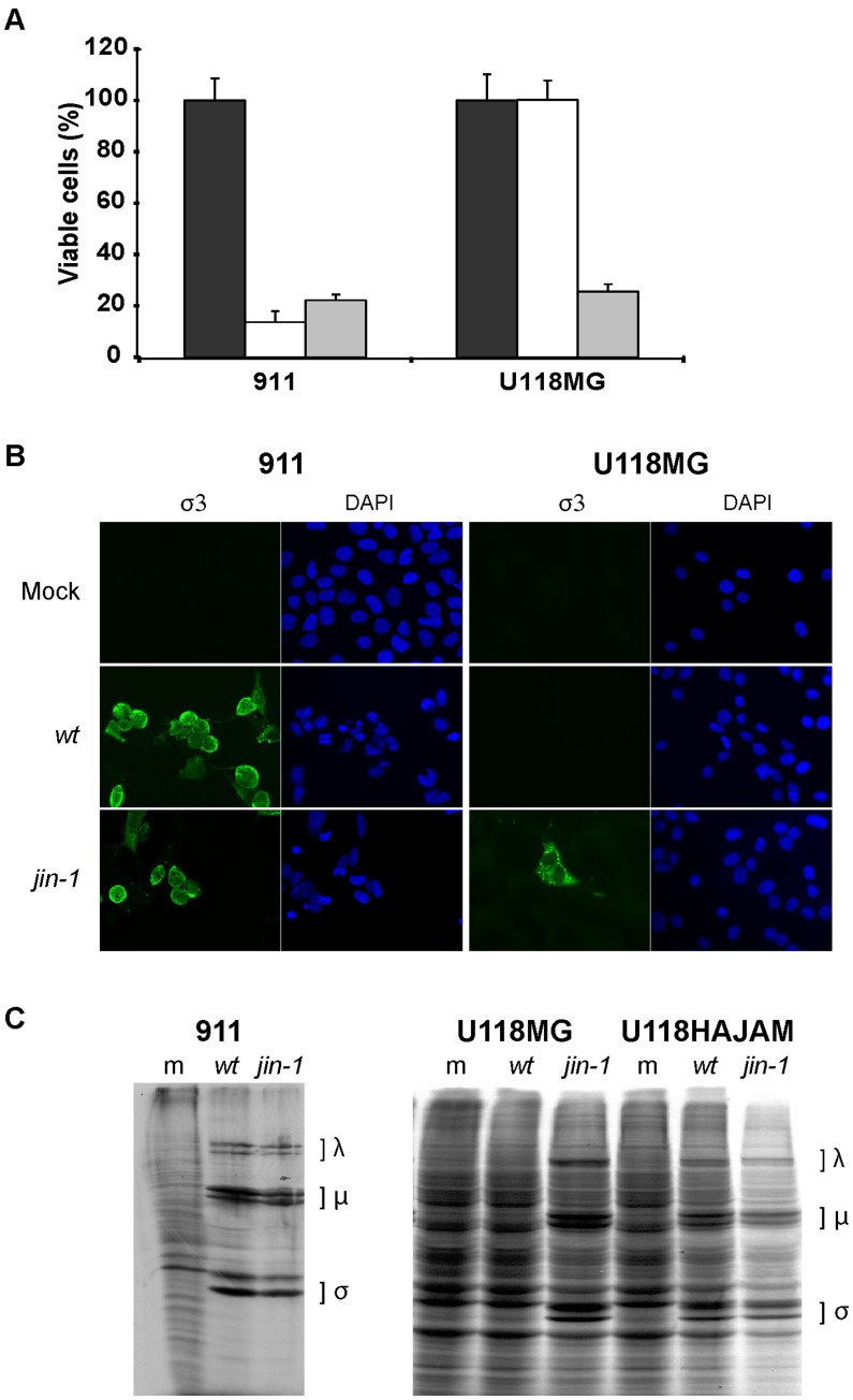
Figure 1. Reovirus mutant jin-1 is able to infect the JAM-A negative cell line U118MG.
(A) Viability assay (WST-1) on 911 and U118MG cells. Cells were mock infected (black bar) or infected with wt T3D (white bar) or jin-1 virus (grey bar) with an MOI of 10, six days post infection. Means (± standard deviation) from three wells. (B) Detection of outer capsid protein σ3 in 911 and U118MG cells after addition of wt T3D or jin-1 virus with an MOI of 5. 40 hr post infection cells were stained with a monoclonal antibody directed against σ3 (4F2) and visualised with a fluorescein isothiocyanate (FITC)-conjugated goat-anti-mouse secondary antibody. The nuclei are visualised with 4′,6-diamidino-2-phenylindole (DAPI). (C) Assessment of reoviral protein synthesis in jin-1 or wt T3D infected cells. Indicated cells were infected with wt T3D or jin-1 virus and labeled with [35S]-methionine once CPE became apparent. 911 cells were infected with an MOI of 1 and U118MG or U118HAJAM cells with MOI of 5. Indicated are the positions of the reoviral λ, µ and σ proteins. m represents mock infected cells; wt: wt T3D infected cells and jin-1: jin-1 infected cells.

