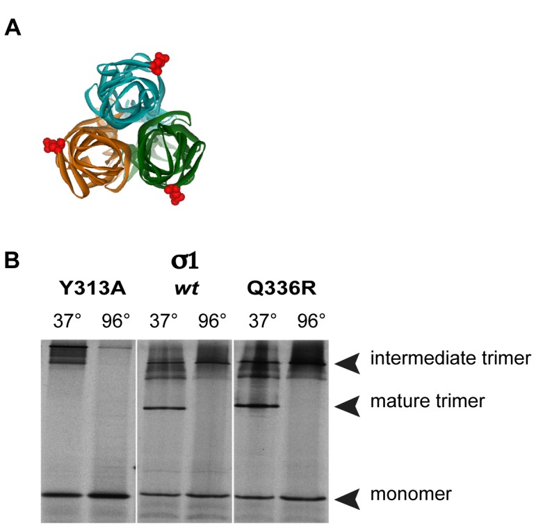
Figure 5. Analysis of Sigma-1 trimers synthesized in vitro.

(A) Top view of σ1-trimer, with colored monomer units (green, turquoise, orange). Position of the Q336R mutation in each monomer is indicated as red CPK symbol (Chappell et al., 2002); PDB ID: 1KKE. The software used for the 3D graphs is Viewerlite 5.0 from Accelrys. (B) [35S] methionine labelled in vitro transcribed and translated products of plasmids pDGC-S1wt (S1wt), pDGC-S1Q336R (S1Q336R) and pDGC-S1Y313A (S1Y313A) were incubated for 30 minutes at 37°C (to stabilize the mature trimers) or boiled for 5 minutes (to disrupt the trimers), before loading on a 10% SDS-polyacrylamide gel at 4°C. The position of the three different conformations is indicated.
