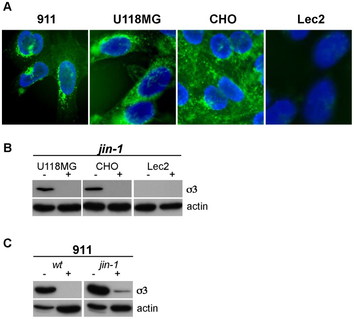
Figure 9. WGA inhibits binding of reovirus to cells.
(A) Detection of Sialic acids in cell lines (911, CHO, U118MG and Lec2) by FITC-labeled WGA immunofluorescence. (B) WGA inhibition of reovirus infection. Prior to exposure of reovirus jin-1, the cells (U118MG, CHO, and Lec2) were mock-treated (−) or treated with 100 µg/ml WGA for 1 hr. at 37°C (+). After exposure of the cells to the virus at 4°C the cells were washed with PBS and incubated for an additional 32 hours in a CO2 incubator before protein lysates were made. For the immunodetection of the σ3 protein the anti-reovirus σ3 (4F2) was used and anti-actin was used as a loading control. (C) WGA inhibition of wt T3D and jin-1 reovirus infection in 911 cells.

