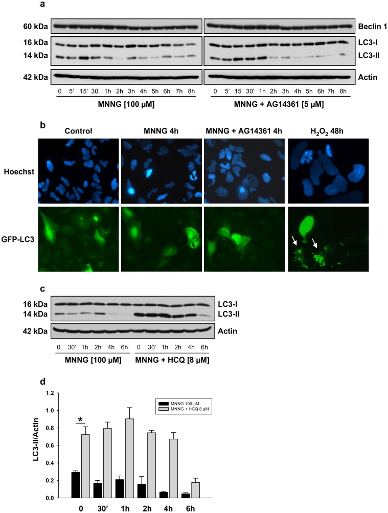
Figure 4. Effect of MNNG exposure on Beclin 1 expression and on the autophagic vesicles marker LC3.
HEK293 cells were treated with 100 µM MNNG alone or in combination with 5 µM AG14361 one hour prior to MNNG exposure. (a) Beclin 1 expression and the conversion of LC3-I to LC3-II were detected by immunoblotting. The blots were also probed with actin antibody to show equal loading. (b) Cells were transiently transfected with the GFP-LC3 plasmid for 24 hours and incubated with MNNG alone or in combination with AG14361. Nuclei were stained with Hoechst 33342. The distribution of GFP-LC3 was examined by fluorescence microscopy 4 hours after the start of MNNG exposure and representative cells were photographed. As a positive control for GFP-LC3 redistribution, cells were treated with 1 mM H2O2 for 48 hours (lower right panel). Punctuated distribution of GFP-LC3 is indicated by the arrows. (c) HEK293 cells were treated with MNNG alone or in combination with the lysosomal protease inhibitor hydrochloroquine sulfate (HCQ) and LC3-II accumulation was detected by immunoblotting. (d) LC3-II/Actin ratios were calculated at each time point. Data are presented as the mean ± standard error of the mean (SEM) of three independent experiments. * P<0.005. One way analysis of variance (ANOVA) shows no significant changes in LC3-II accumulation in MNNG+HCQ-treated cells between 0 and 4 hours.

