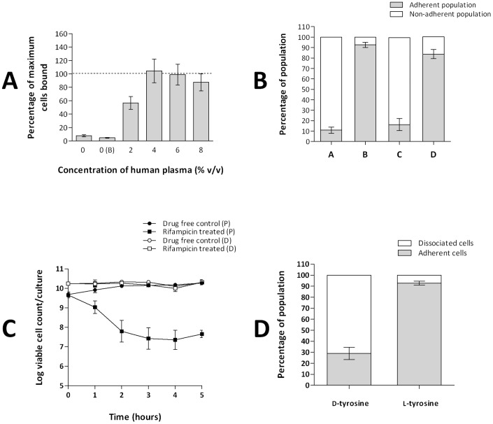
Figure 1. Development and validation of the cellulose disk static biofilm model.
A. Effect of human plasma on the adherence of S. aureus SH1000 to cellulose disks. On the x-axis, 0 represents no human plasma or buffer and 0(B) is buffer only. Data indicate the proportion of adherent cells, in 48 hr disk cultures, as a percentage of the maximum achieved at 10% (v/v) human plasma (dashed line) (1.8×10¹⁰ cfu/disk). Data from three experimental replicates. Error bars indicate standard error. B. Determination of the proportion of adherent and planktonic cells present in S. aureus SH1000 cellulose disk cultures. (A) Cultures grown for 48 hrs in the absence of human plasma. (B) Cultures grown for 48 hrs with human plasma (4% v/v) added prior to inoculation. (C) Cultures grown for 144 hrs with human plasma (4% v/v) added prior to inoculation. (D) Cultures grown for 144 hrs with human plasma (4% v/v) added prior to inoculation and every 48 hrs. Data from three experimental replicates. Error bars indicate standard error. C. Time-kill curves of S. aureus SH1000 exponential phase planktonic (P) and 48 hr cellulose disk (D) cultures exposed to 0.25 mg/L rifampicin. Data from three experimental replicates. Error bars indicate standard deviations. D. The dissociation of adherent cells of S. aureus SH1000 from cellulose disk cultures in the presence of d-tyrosine (100 µM) and l-tyrosine (100 µM). Data from three experimental replicates. Error bars indicate standard error. Methodology and additional information regarding validation of the cellulose disk model can be found in Supporting Information S1.

