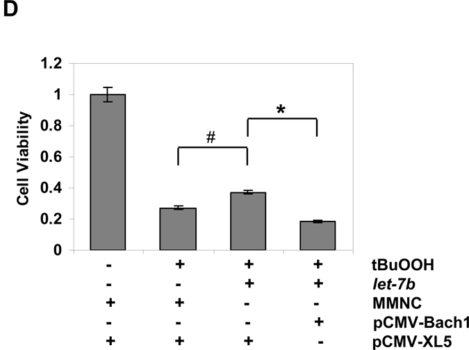
Figure 6. Over-expression of let-7b protects cells from oxidative injury in Huh-7 cells.
Huh-7 cells were transfected for 48 h with 50 nM of let-7b or MMNC by Lipofectamine 2000. Cells were then incubated with the indicated concentrations of tBuOOH for 4 or 6 h. CellTiter-Glo® Reagent was added for CellTiter-Glo luminescent cell viability assay on a Synergy HT microplate reader with integration time set for 0.25 to 1 second. Decreases in luminescence were taken as an index of cellular cytotoxicity. Data represent means ± SE of triplicate determinations. Data are presented as means ± SE, n=3. * differs from MMNC only, P<0.05. Susceptibility of Huh-7 cells transfected with MMNC and let-7b to tBuOOH induced oxidative injury was compared. (A) 4 h after tBuOOH treatment (*differs from MMNC, p= 0.03); (B) 6 h after tBuOOH treatment (*differs from MMNC, p=0.03); (C) WB analysis of over-expression of Bach1 by pCMV-Bach1; (D) The protection of cells by let-7b against oxidative injury was ablated following over-expression of Bach1. Huh-7 cells were transfected with 0.4 µg/mL of pCMV-Bach1 or pCMV-XL5, a control vector without Bach1 cDNA insertion for 24 h, and then transfected with 50 nM of let-7b or MMNC for 48 h. Cells were then incubated with 400 µM of tBuOOH for 6 h. Cell viability assays were performed as described above. Data are presented as means ± SE, n=3. #p=0.006 and *p=0.001.


