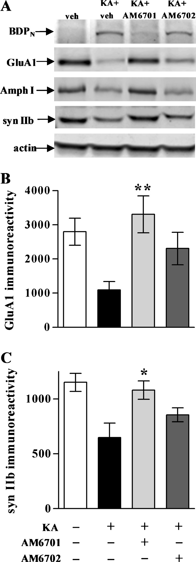
Fig. 7.
Synaptic and cytoskeletal protection in the kainic acid (KA) rat model. No-insult control rats from Figs. 5 and 6, along with the KA groups treated with or without carbamate inhibitor, were subsequently assessed by immunoblot for spectrin breakdown product (BDPN), GluA1, amphiphysin I (Amph I), synapsin IIb (syn IIb), and actin in dissected hippocampal tissue (a). Integrated optical densities for GluA1 (b) (analysis of variance, p < 0.01; n = 7–9 per group) and Amph I (C) (p < 0.01) are shown as means ± SEM, indicating that AM6701 is more protective than AM6702 against excitotoxin-induced synaptic marker decline. Post hoc tests compared to KA only group: *p < 0.05, **p < 0.01

