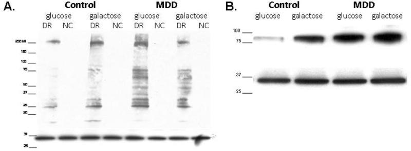
Figure 1.
A) Representative DNPH blot. Western blot probed with DNPH antibody (top) and re-probed with GAPDH antibody (bottom). Blot shows one control-MDD matched pair, derivatized reaction (DR) and negative control (NC) for each sample with molecular weight marker; from left to right lanes read: control in glucose medium, control in galactose medium for 24 hours, MDD in glucose medium, and MDD in galactose medium for 24 hours. Directly below the DNPH blot is the GAPDH restain of the same PVDF membrane displaying equal protein loading. B) Representative GR blot. Western blot probed with Glutathione Reductase antibody (top) and re-probed with GAPDH antibody (bottom). Blot shows one control-MDD matched pair with molecular weight marker; from left to right lanes read: control in glucose medium, control in galactose medium, MDD in glucose medium, and MDD in galactose medium. Directly below the GR blot is the GAPDH restain of the same PVDF membrane displaying equal protein loading.

