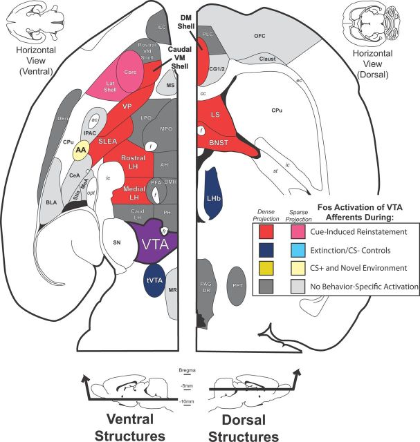
Figure 11.
Summary of behavior-related Fos activation of VTA afferents. A summary of the VTA inputs that were Fos activated during cue-induced reinstatement and other behaviors are diagrammed in the horizontal plane (modified from Paxinos and Watson, 2007), with afferents lying ventrally in the brain shown at left (plane shown ∼7.8 mm ventral of bregma), and dorsal structures shown at right (plane shown ∼4.6 mm ventral of bregma). Structures labeled in red provided relatively dense inputs to VTA that expressed more Fos in cue-induced reinstatement animals than any of the control groups. Pink structures provided relatively sparse afferents that were activated most in reinstatement animals. Dark blue structures provided relatively dense VTA afferents, and that expressed more Fos in animals in which cocaine was not expected (extinction and CS− groups) than in other groups. AA (light yellow) provided a relatively sparse input to VTA that was Fos-activated in reinstatement and locomotor control animals, indicating possible involvement in behaviorally activating situations, but not cocaine-seeking in particular. Gray structures provided inputs to VTA (dark gray, relatively dense inputs; light gray, relatively sparse inputs) that did not express Fos preferentially in any behavioral condition. VTA is colored in purple. AH, Anterior hypothalamus; Caudal LH, LH caudal of bregma −3.5 mm; Caudal VM Shell, ventromedial nucleus accumbens shell caudal of bregma +1.5 mm; Core, nucleus accumbens core; CPu, caudate–putamen; DEn, dorsal endopiriform nucleus; DM Shell, dorsomedial accumbens shell; Lat Shell, lateral nucleus accumbens shell; LPO, lateral preoptic area; MeA, medial amygdala; MS, medial septum; MPO, medial preoptic area; MR, medial raphe nucleus; OFC, orbitofrontal cortex; PAG/DR, periaqueductal gray/dorsal raphe nucleus; PH, posterior hypothalamus; PLC, prelimbic cortex; PPT, pedunculopontine nucleus; Rostral LH, LH rostral of bregma −2.5 mm; Rostral VM Shell, ventromedial nucleus accumbens shell rostral of bregma +1.5 mm; SN, substantia nigra; Stia, intra-amygdaloid stria terminalis. White matter tracts are labeled in italics: opt, optic tract; ac, anterior commissure; f, fornix; ic, internal capsule; ec, external capsule; st, stria terminalis.

