
Figure 3.
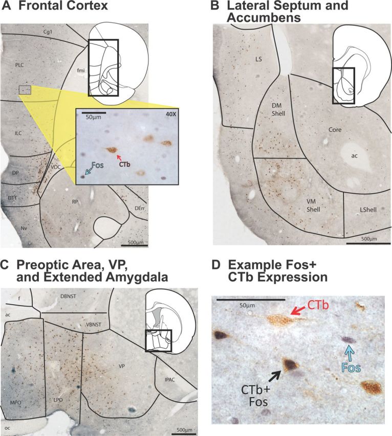
Pattern of major projections to VTA from telencephalon. Representative examples are shown of several regions in which significant numbers of VTA-projecting neurons were observed after VTA CTb injections. Background photographs were taken at 10× magnification and compiled into mosaics to show regional variations in CTb staining patterns (scale bars shown in each panel). Insets show corresponding atlas pages modified from Paxinos and Watson (2007), and higher-magnification photographs of CTb/Fos staining. A, Many CTb+ neurons were seen in ventral portions of medial prefrontal cortex. Inset shows a close up (40× magnification) of Fos+ and CTb+ neurons in ventral prelimbic cortex. B, Many CTb+ neurons were seen in medial accumbens shell and lateral septum. C, Many CTb+ neurons were seen in a band stretching from the preoptic area laterally into ventral BNST and into ventral pallidum. D, A 60× magnification photo of a Fos+ neuron (blue arrow), a CTb+ neuron (red arrow), and a CTb/Fos costained neuron (black arrow) are shown, taken from a section within ventral pallidum. Image was merged from a z-stack using ImageJ (NIH) software. ac, Anterior commissure; Core, nucleus accumbens core, DBNST, dorsal BNST, DEn, dorsal endopiriform cortex; DM Shell, dorsomedial nucleus accumbens shell; DP, dorsal peduncular cortex; DTT, dorsal tenia tecta; f, fornix; fmi, forceps minor of the corpus callosum; ILC, infralimbic cortex; LPO, lateral preoptic area; LShell, lateral nucleus accumbens shell; MPO, medial preoptic area; NvN, navicular nucleus; oc, optic chiasm; PLC, prelimbic cortex; RP, rostral pole of the nucleus accumbens; VBNST, ventral BNST; VM Shell, ventromedial nucleus accumbens shell; VOC, ventral orbitofrontal cortex.