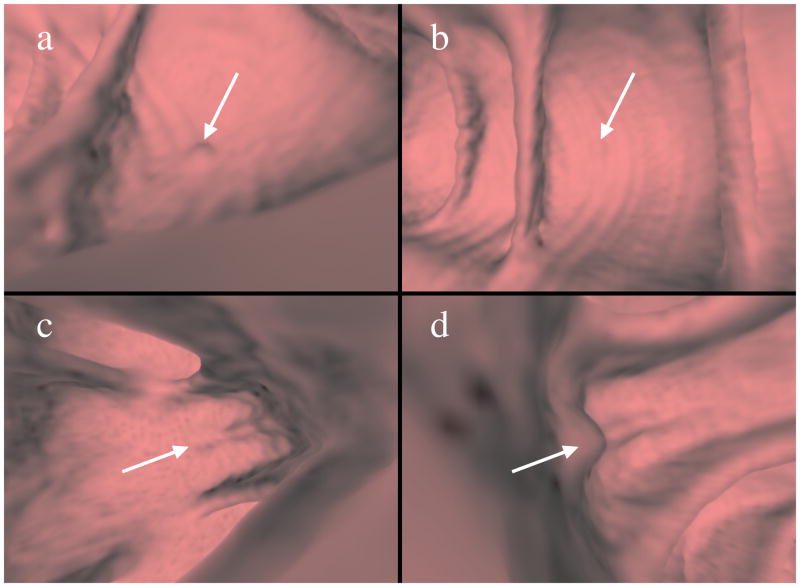
Fig. 1.
Need for instance ranking. Images (a) and (b) are taken from a positive bag that illustrates a 6 mm sessile polyp, while images (c) and (d) are from a negative bag showing a false positive at the base of a fold. Images (a) and (c) show instances of the detections that should be preferred as they are more representative of their respective classes. Instances in (b) and (d) appear misleading and could lead to incorrect classification.

