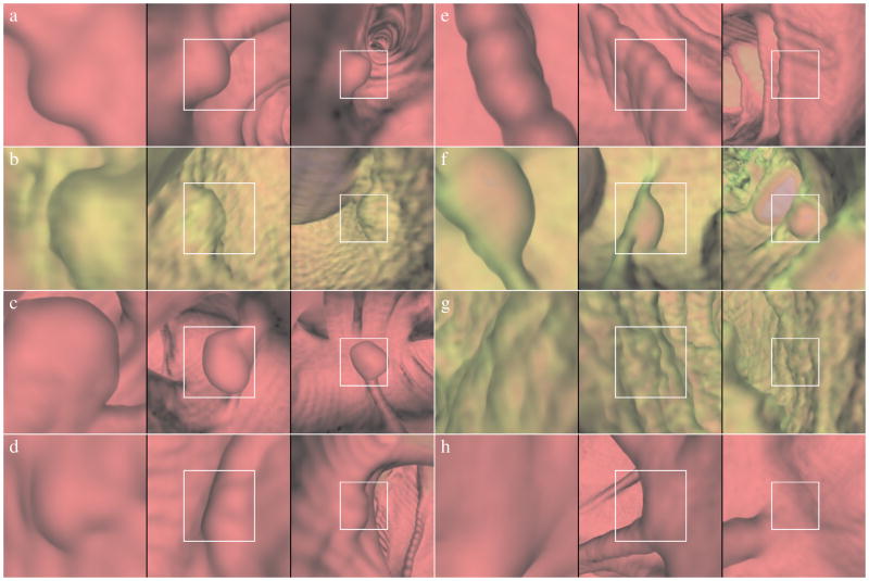
Fig. 3.
Representative frames extracted from typical videos with different viewpoints and viewing angles. Images (a)–(d) show true polyps. Image (a) shows a 6 mm sessile polyp, (b) a 1.1 cm sessile polyp, (c) a 1.1 cm pedunculated polyp, and (d) a 6 mm sessile polyp. Images (e)–(h) show common false positives. Image (e) shows a detection on a haustral fold, (f) an air bubble false positive, (g) a detection arising from rough texture, and (h) a detection on a taenia coli. The green color in (b), (f), and (g) demonstrates that the detection is covered in iodinated endoluminal contrast material.

