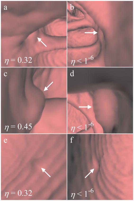
Fig. 9.
Ranked instances of flat polyps. The left column and right column show the highest- and lowest-rated instance for each detection, respectively. (a)–(b) 6 mm flat polyp, (c)–(d) 8 mm flat polyp, (e)–(f) 6 mm flat polyp. The polyps in (a)–(b) and (e)–(f) each had scores in the bottom 20% of all true polyps. The polyp in (c)–(d) had a score in the upper third of all true polyps.

