
Figure 1.
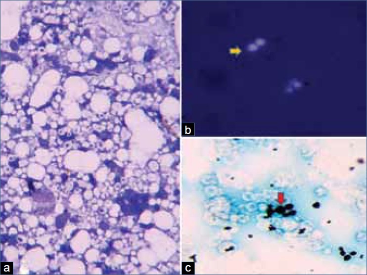
(a) Numerous spherical yeast cells of variable size with occasional budding forms surrounded by a clear halo suggestive of a capsule, lymphocytes and histiocytes in a necrotic background (MGG, ×200).(b) Budding yeast forms surrounded by a thick capsule (yellow arrow) (India ink preparation, ×400). (c) Budding yeast forms (red arrow) of variable size surrounded by a clear halo (Gomori's methenamine silver, ×400)