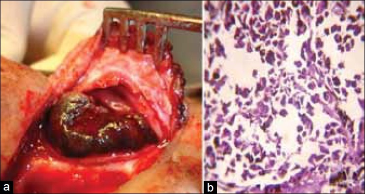
Figure 2.

(a) Intraoperative tumor-mass before resection. (b) Section showed medium to large size epithelioid and spindle cells arranged in lobules and small bundles with pleomorphic, hyperchromatic nuclei with prominent nucleoli. Both extra and intracytoplasmic melanin pigment was noted (H and E, ×400)
