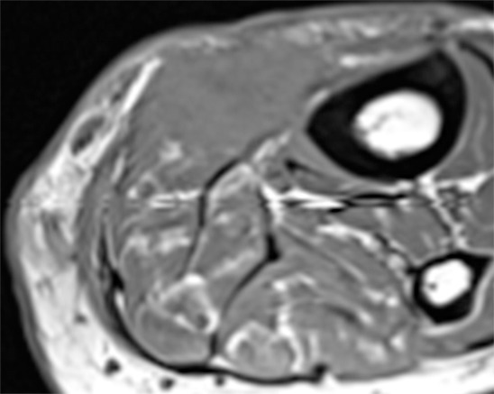
Figure 7b:

67-year-old woman with recurrent malignant fibrous histiocytoma. The advantages of functional techniques over anatomic imaging are highlighted. (a) Axial fat-suppressed T2 weighted image (TR/TE 3560/64) shows a relatively low to intermediate signal area of signal abnormality in the surgical bed (rectangle) with surrounding postoperative inflammation. (b) Axial T1 weighted image (TR/TE 580/20) shows architectural distortion suspicious for a recurrent mass. (c) ADC map shows a low signal intensity region with ADC value of 0.4, highly suspicious for recurrent tumor. (d) Finally, a contrast-enhanced coronal view from a DCE-MR imaging study (shown here at 20 seconds) shows the neovascularity of the recurrent tumor to best advantage.
