Figure 1.

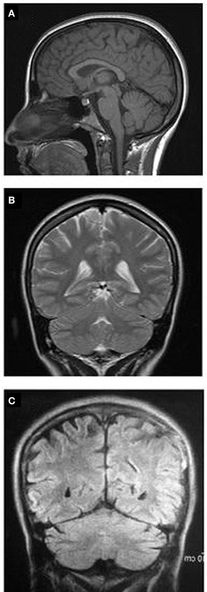
MRI images showing sagittal (A) and coronal (B) sections of case 1 with facultative QL; (C) coronal MRI of case 2 with a paralyzed leg. Notice the entirely normal brain structures in both cases.

MRI images showing sagittal (A) and coronal (B) sections of case 1 with facultative QL; (C) coronal MRI of case 2 with a paralyzed leg. Notice the entirely normal brain structures in both cases.