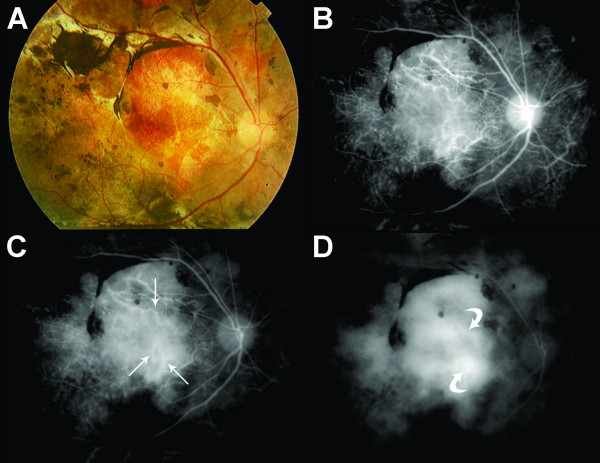
Figure 1.
Color fundus photography (A) as well as early, mid and late phase indocyanine green angiography (B,C,D, respectively) from a representative patient with Vogt-Koyanagi-Harada and long-standing disease. Note “fuzzy vessels” (arrows) on early and mid phases ICGA, and “late diffuse hyperfluorescence” (curved arrows) on late phase of the exam.

