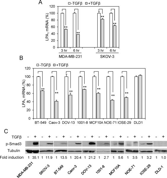
Figure 1. TGFβ inhibits expression of LPA1 mRNA.
A. MDA-MB-231 and SKOV-3 cells were cultured with TGFβ (2.5 ng/ml) or vehicle for 3 and 6 hours. LPA1 mRNA levels were determined by RT and qPCR. The mRNA levels of LPA1 in TGFβ treated cells were presented as percentages relative to those in control cells (defined as 100%). B. Multiple cancer cell lines, immortalized breast (MCF-10A) and ovarian (IOSE-29) epithelial cell lines, primary mammary (1001-8) and ovarian (NOE71) epithelial cells were treated for 6 hours with TGFβ (2.5 ng/ml) and analyzed for LPA1 mRNA expression as in A. C. Cancer cell lines and primary cells were treated with TGFβ (2.5 ng/ml) or vehicle for 1 hour before lysis with SDS sample buffer and immunoblotting analysis of Smad3 phosphorylated at Ser423/425. The intensity of phospho-Smad3 in each cell line was quantified by densitometry and presented as fold of that in control cells (arbitrary 1.0).

