
Figure 5.
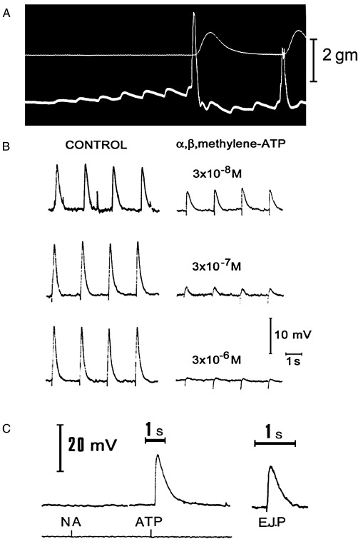
(A) Excitatory junction potentials in response to repetitive stimulation of adrenergic nerves (white dots) in the guinea pig vas deferens. The upper trace records the tension, the lower trace the electrical activity of the muscle recorded extracellularly by the sucrose gap method. Note both summation and facilitation of successive junction potentials. At a critical depolarization threshold, an action potential is initiated which results in contraction. [From Burnstock and Costa (1975), reproduced with permission of Chapman and Hall.] (B) The effect of various concentrations of α,β-methylene ATP (α,β-meATP) on excitatory junction potentials (EJPs) recorded from the guinea pig vas deferens (intracellular recording). The control responses to stimulation of the motor nerves at 0.5 Hz are shown on the left. After at least 10 min in the continuous presence of the indicated concentration of α,β-meATP, EJPs were recorded using the same stimulation parameters. The EJPs are clearly reduced in magnitude in the presence of α,β-meATP. Notice also that in the control cells, several large spontaneous EJPs were seen, whereas after α,β-meATP, no spontaneous EJPs were recorded. [Reproduced from Sneddon and Burnstock (1984a), with permission of Elsevier.] (C) Spritzed ATP, but not NA, mimicked the EJP recorded in the vas deferens. [Reproduced from Burnstock and Verkhratsky (2012) (Book Chapter 6) with permission of Springer.]