
Figure 13.
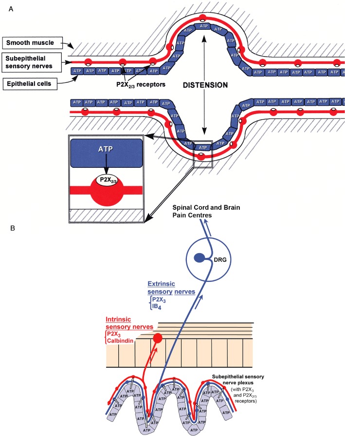
Purinergic mechanosensory transduction. (A) Schematic representation of hypothesis for purinergic mechanosensory transduction in tubes (e.g. ureter, vagina, salivary and bile ducts, gut) and sacs (e.g. urinary and gall bladders, and lung). It is proposed that distension leads to release of ATP from epithelium lining the tube or sac, which then acts on P2X3 and/or P2X2/3 receptors on subepithelial sensory nerves to convey sensory/nociceptive information to the CNS. [From Burnstock (1999), reproduced with permission from Blackwell Publishing.] (B) Schematic of a novel hypothesis about purinergic mechanosensory transduction in the gut. It is proposed that ATP released from mucosal epithelial cells during moderate distension acts preferentially on P2X3 and/or P2X2/3 receptors on low-threshold subepithelial intrinsic sensory nerve fibres (labelled with calbindin) to modulate peristaltic reflexes. ATP released during extreme (colic) distension also acts on P2X3 and/or P2X2/3 receptors on high-threshold extrinsic sensory nerve fibres [labelled with isolectin B4 (IB4) ] that send messages via the dorsal root ganglia (DRG) to pain centres in the central nervous system. [From Burnstock (1999), reproduced with permission from Wiley.]