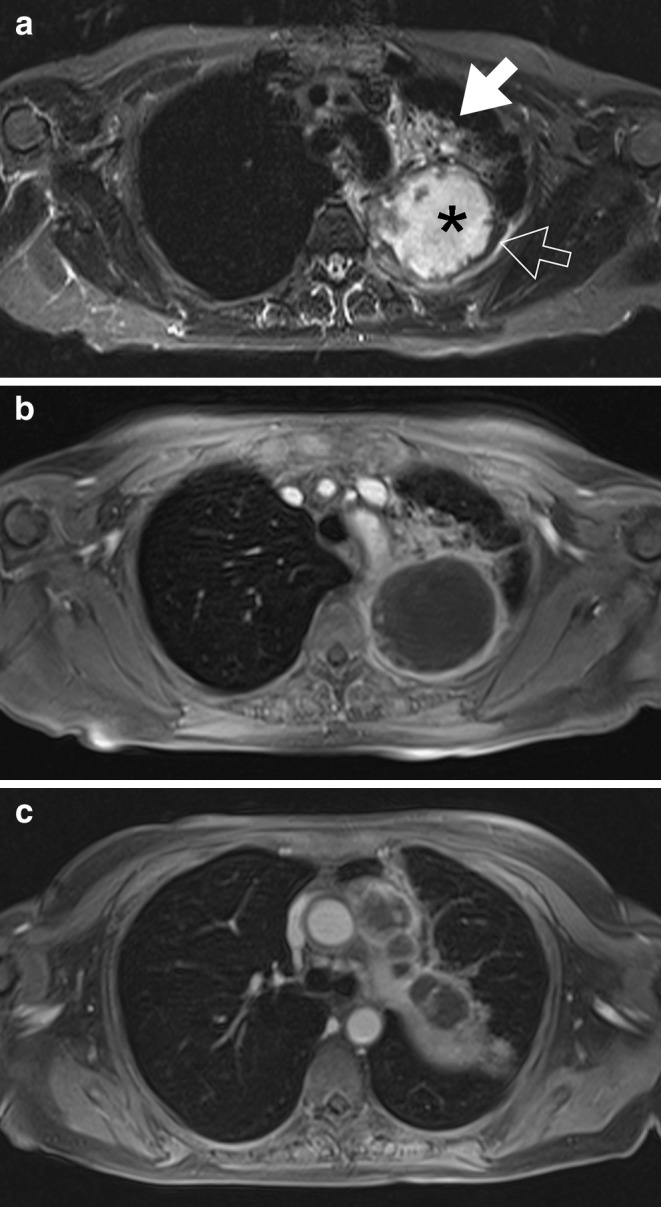
Fig. 3.

A 56-year-old female patient with small cell lung cancer. The transverse T2-weighted fat-saturated (a) and T1-weighted contrast-enhanced fat-saturated 3D-GRE images (b, c) show a large, centrally necrotic mass in the left upper lobe with large peri-hilar lymph node metastases. Note the high soft tissue contrast between alelectatic lung (open arrow), small rim of solid tumour (filled arrow) and colliquated central portion of the mass (asterisk)
