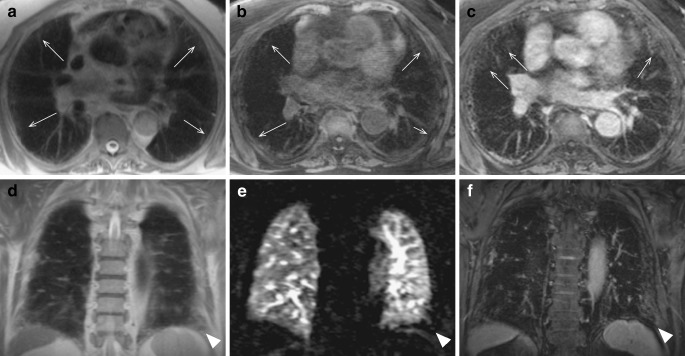
Fig. 6.
Subtle subpleural reticulation in a patient with fibrotic-predominant NSIP. The interlobular reticulation (thin arrows) is more evident after contrast administration (c and f). A perfusion defect (arrowhead in e) is associated to the peripheral fibrotic changes at the left lateral costo-phrenic angle (arrowheads in d and f)

