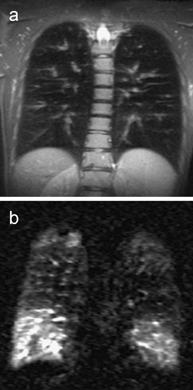
Fig. 4.
An 18-year-old male cystic fibrosis patient, coronal T2-weighted half Fourier fast spin echo sequence (a) and coronal subtraction perfusion image (b). Notice the severe mucus plugging in the morphological T2-weighted image. The subtraction perfusion image shows correspoding areas with perfusion loss due to hypoxic vasoconstriction. Due to redistribution of perfusion both lower lobes show a high perfusion signal

