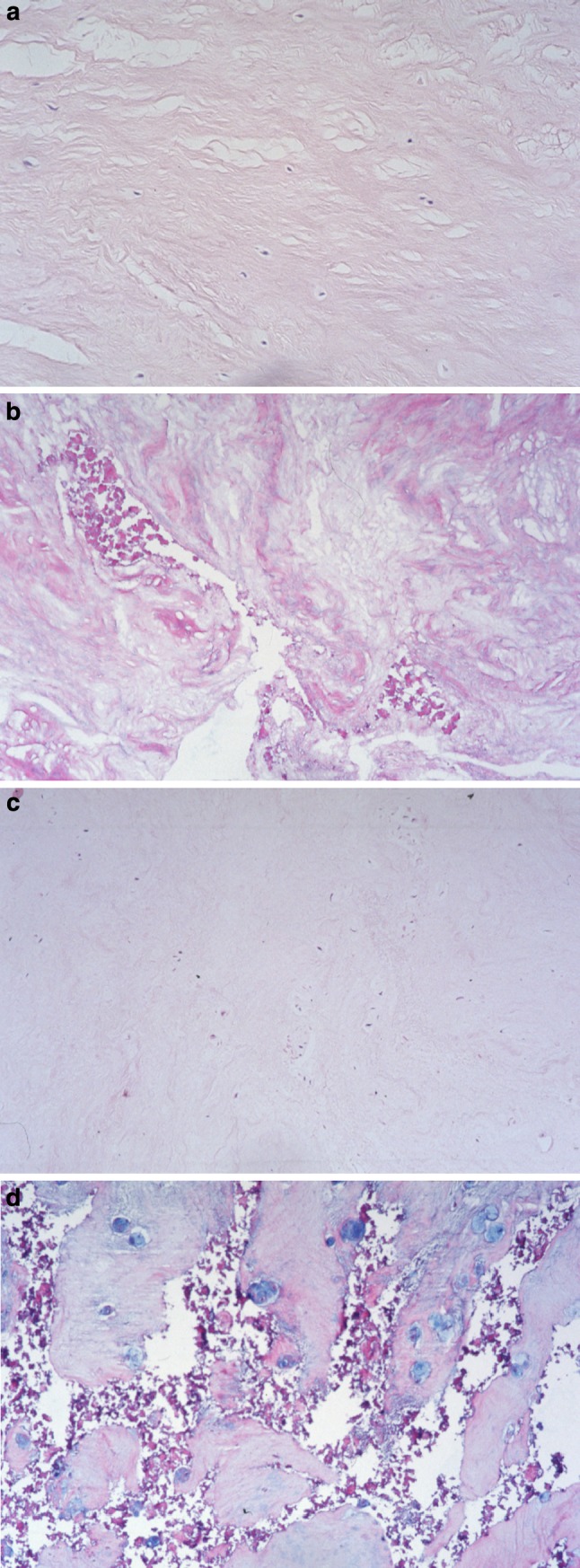
Fig. 2.

Histomorphological features of normal and pathological disc tissue specimens [Haematoxylin and Eosin (H&E), ×300]. a Sample from the (inner) annulus fibrosus with fairly parallel arranged collagen fibres and few unremarkable cells (HDS score 0). b Annulus fibrosus tissue area with clefting and extensive granular matrix alteration (HDS score 6). c Nucleus pulposus section with fibrocartilaginous matrix and very slightly enhanced, single layered nuclear chondrocytes (HDS score 1). d Nucleus pulposus section with extensive clefting, granular matrix changes and focal clonal chondrocyte proliferation (HDS score 10)
