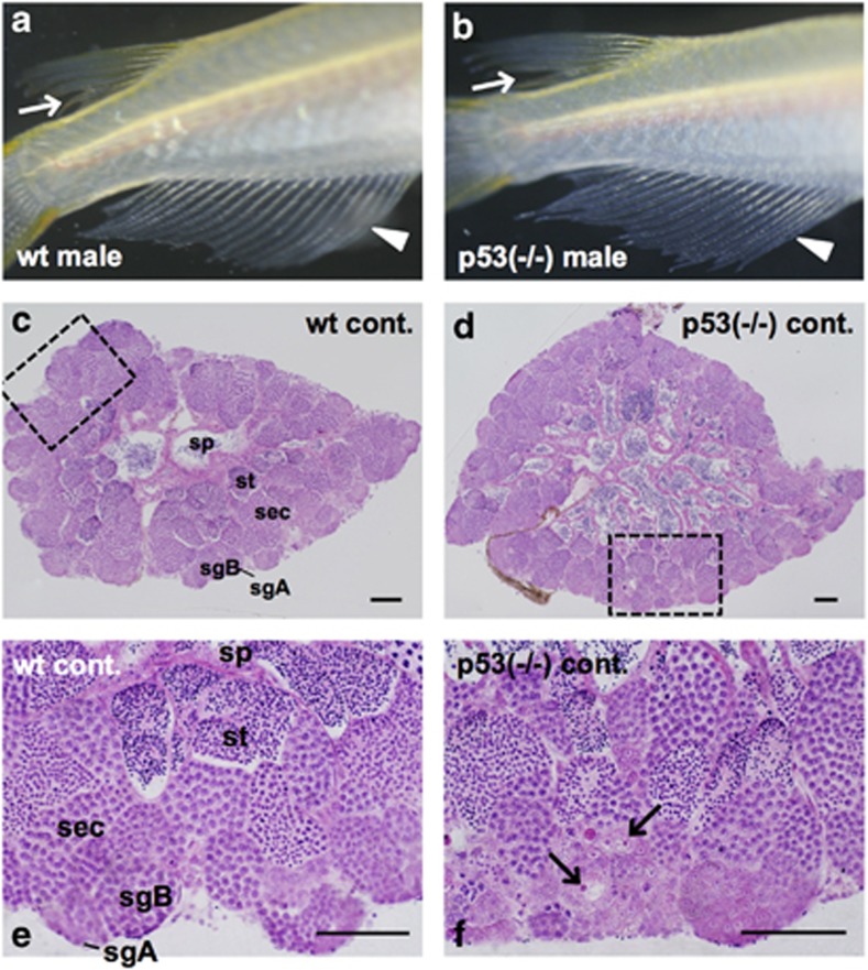
Figure 1.
Testis–ova were differentiating spontaneously in the non-irradiated p53(−/−) testis. Representative images of the secondary sex characteristics of p53(−/−) medaka adults that are genetically male (b), showing typical male-type external appearance of wt male medaka (a) such as rougher edges of dorsal fins (arrows in a and b) and sharply long anal fins (arrowheads in a and b). H&E-stained sections of non-irradiated testes of wt and p53(−/−) fishes at 6 months old. (c) wt testis. (d) p53(−/−) testis. (e) Enlarged view of the boxed area in (c). (f) Enlarged view of the boxed area in (d) showing a small number of characteristic cells positioned in the cysts with type A or B spermatogonia, of which the nucleolus was strongly H&E-stained and the nucleus was faintly stained (arrows in f). sgA, type A spermatogonia; sgB, type B spermatogonia; sec, spermatocytes; st, spermatids; sp, spermatozoa. Scale bars represent 50 μm

