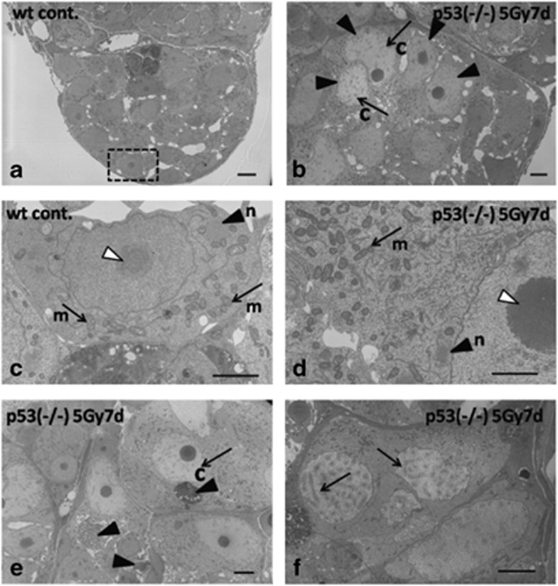
Figure 4.
Electron microscopic observations of testis–ova in p53(−/−) mutants 7 days after irradiation with γ-rays (5 Gy). Morphological changes of testis–ova after γ-ray (5 Gy) irradiation. (a) Electron microscopic observation of wt spermatogonia. (c) Enlarged view of the boxed area in (a) showing the germinal dense body (nuage) in the cytoplasm (arrowhead with n in c), which is closely associated with large aggregations of mitochondria (arrows with m in c). The nucleolus of a spermatogonia is shown by an open arrowhead in c. (d) In the cytoplasm of p53(−/−) testis–ova, larger and more electron-dense nucleoli (e.g., open arrowhead in d) are present. It is noticeable that an extensive number of mitochondria and more electron-dense nuage are present (arrow with m and arrowhead with n in d) compared with those of wt spermatogonia (arrows with m and arrowhead with n in c). (b) The testis–ova in the irradiated testis increased synchronously (arrowheads in b) and they had a characteristic appearance of short and thick chromatin (arrows with c in b and e). (e) Apoptotic condensed nuclei were observed nearby testis–ova (arrowheads in e). (f) Some testis–ova have synaptonemal complexes in the nucleus (arrows in f). Chromatin (c), mitochondria (m), nuage (n). Scale bars represent 2 μm in c and d; 5 μm in a, b, e and f

