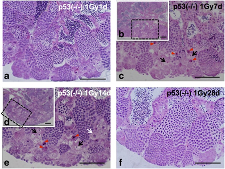
Figure 7.
Histological changes of testis–ova in p53(−/−) testes during 1 month after γ-ray (1 Gy) irradiation. (a) H&E-stained section of p53(−/−) testis 1day after irradiation showing no apparent histological changes. (b) Histological H&E-stained section of p53(−/−) testis 7 days after irradiation and (c) enlarged view of the boxed area in B, showing pyknotic cells (arrowheads in c) and increased testis–ova (arrows in c). (d) Histological H&E-stained section of p53(−/−) testis 14 days after irradiation and (e) enlarged view of the boxed area in (d), showing pyknotic cells (arrowheads in e) and testis–ova (arrows in e) were present. (f) Histological H&E-stained section of p53(−/−) testis 28 days after irradiation showing that spermatogenesis was restored almost completely, and that the number of testis–ova had decreased. Scale bars represent 50 μm

