Figure 8.
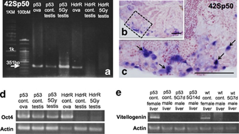
Gene expressions in p53(−/−) testes after γ-ray (5 Gy) irradiation. (a) Transcript of 42Sp50 were amplified both in non-irradiated and irradiated p53(−/−) testes with γ-rays (5 Gy), as well as in p53(−/−) and wt Hd-rR ovaries, while not amplified in non-irradiated and irradiated wt testes. (b) Expression of 42Sp50 in irradiated p53(−/−) testes was investigated on histological sections by ISH and as clearly shown in the enlarged view (c) of the boxed area in b, testis–ova in the p53(−/−)-irradiated testes were positive (blue-stained cells; arrows in c). (d) Transcript of Oct4 were amplified both in non-irradiated and irradiated p53(−/−) testes with γ-rays (5 Gy), as well as in p53(−/−) and wt Hd-rR ovaries, while not amplified in non-irradiated and irradiated wt testes. (e) Transcripts of vitellogenin were not amplified from male liver cDNA of wt and p53(−/−), without irradiation and with γ-ray (5 Gy) irradiation at 7days and 14 days. However, they were clearly amplified from female liver of both control wt and p53(−/−). Scale bars represent 50 μm

