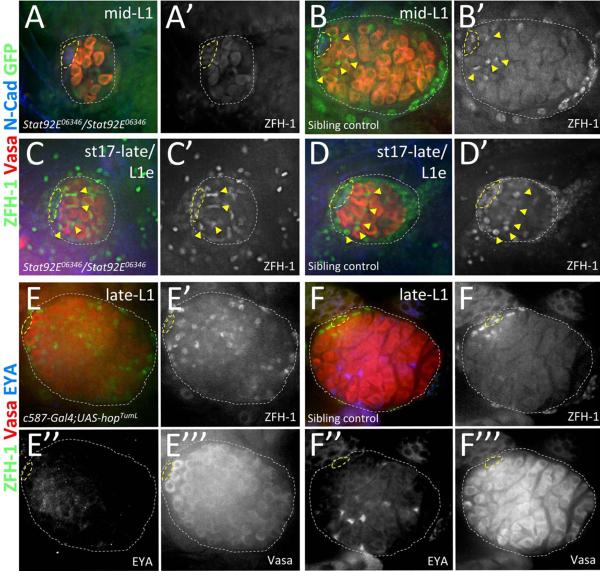
Figure 4. Jak-STAT signaling is necessary and sufficient for CySC maintenance in larval testes.
Late-stage embryos and 1st instar larval testes immunostained with anti-Vasa (A–F, red; E'”& F'” alone), anti-ZFH-1 (A–F, green; A'–D', in black and white with GFP; E' & F' alone), and either anti-GFP (A–D, green; A' & B' in black and white with ZFH-1) to determine genotype of Stat92E06346 mutants (see methods) or anti-EYA (E & F, blue; E” & F” alone) or. All images with testis anterior oriented left. Hub (yellow dashed lines) and testes (white dotted lines) outlined. [A, B] mid-L1 testes from (A) a Stat92E06346 homozygous mutant lacking high-level ZFH-1 expression in somatic cells around the hub and from (B) a sibling controls with ZFH-1 enriched in hub-adjacent cells (yellow arrowheads). [C, D] stage 17-late/L1 early testes from (A) a Stat92E06346 homozygous mutant and (B) a sibling control both showing high-level ZFH-1 in somatic cells adjacent to the hub (yellow arrowheads). [E, F] Late-L1 testes with Jak hyper-activated in the somatic gonad (E; c587-Gal4/+;UAS-hopTumL/+) show expanded expression of high-level ZFH-1 and no EYA expression in cyst cells, while age-matched sibling controls (F; c587-Gal4/+;CyO/+) show high-level ZFH-1 restricted to hub-adjacent CySCs and EYA expression in somatic nuclei interspersed throughout the testis posterior.

