
Figure 2.
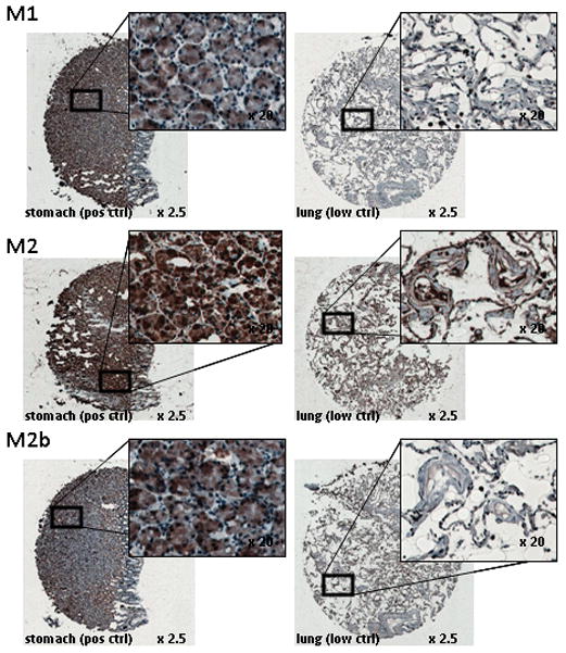
Examples of RNR M1, M2, and M2b antibody staining. IHC staining on FFPE stomach crypt cells for RNR M1, M2, and M2b is intense (3+) in the cytoplasm and nucleus. In contrast, lung pneumocytes show very low (0) stain intensity for RNR M1 and M2b and low (1=) stain intensity for RNR M2.