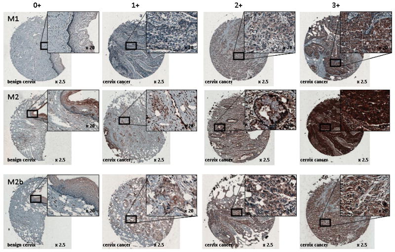
Figure 3.

RNR M1, M2, and M2b immunoreactivity in uterine cervix benign and cancer tissues. Columns indicated brown color staining intensity 0 (< 5% of cells), 1 (5% to < 25%), 2 (25% to < 75%), and 3 (≥ 75%) scale. Rows identify RNR subunit. Magnification is indicated.
