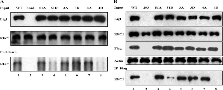
FIGURE 2.
Interaction of hLigI phosphomutants with RFC. A, purified RFC complex (1 μg) was incubated with wild-type hLigI and the indicated phosphomutants (1 μg of each) prior to immunoprecipitation with hLigI antibody (2). Lane 1, wild type hLigI; lane 2, no protein; lane 3, hLig 51A; lane 4, hLigI51D; lane 5, hLigI3A; lane 6, hLigI3D; lane 7, hLigI4A; lane 8, hLigI4D. hLigI (LigI) and RFC p140 (RFC1) were detected by immunoblotting. Upper panel, 10% of the hLigI input. Center panel, 10% of RFC input. Bottom panel, immunoprecipitated RFC p140. B, immunoprecipitation of extracts from 293 cells (4 × 10⁸ cells) expressing the following: lane 1, FLAG-tagged wild-type hLigI; lane 2, no FLAG-tagged protein; lane 3, FLAG-tagged hLigI51A; lane 4, FLAG-tagged hLigI51D; lane 5, FLAG-tagged hLigI3A; lane 6, FLAG-tagged hLigI3D; lane 7, FLAG-tagged hLigI4A; lane 8, FLAG-tagged hLigI4D. The levels of endogenous and FLAG-tagged hLigI (LigI and FLAG), β-actin (Actin), and RFC p140 (RFC1) in the extracts (Input, 5%) were determined by immunoblotting with the indicated antibodies. The extracts were incubated with the FLAG antibody (IP FLAG), and the immunoprecipitates were probed for RFC p140 (RFC1) by immunoblotting.

