
FIGURE 5.
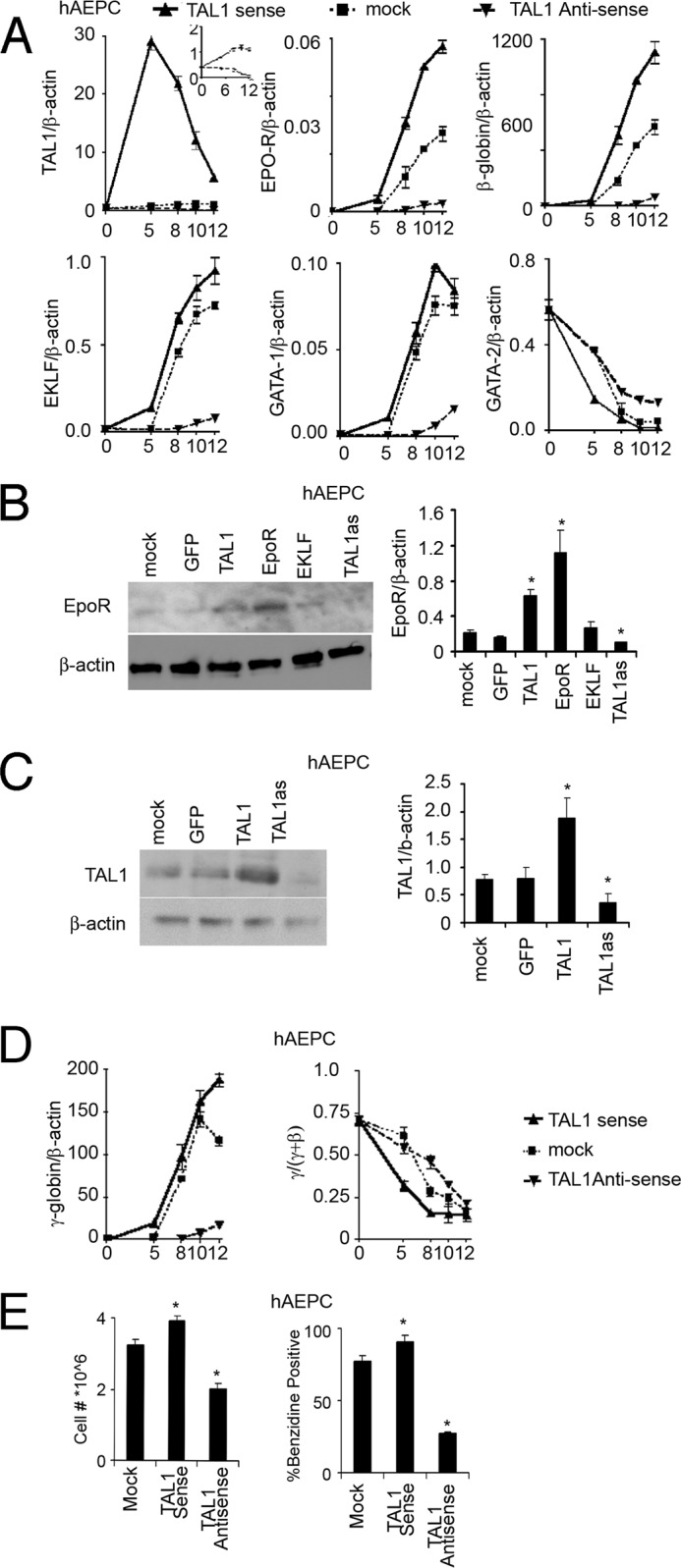
Forced expression of TAL1 in human primary erythroid progenitor cells. A, expression of endogenous TAL1, EPO-R, β-globin, EKLF, GATA-1, and GATA-2 were monitored during 12 days of EPO stimulation with TAL1 sense (triangle, solid line), antisense (inverted triangle, dashed line), and mock (square, dotted line) forced expression in hAEPC. Results are normalized to β-actin expression. TAL1 antisense and mock transfection efficiency was shown in the inset panel for clarity. B, increase in EPO-R protein expression by overexpression of GFP, TAL1, EPO-R, and EKLF and TAL1 knockdown (TAL1 as) are shown in hAEPC using Western blotting with β-actin as loading control. C, increase in TAL1 protein expression by GFP and TAL1 overexpression and TAL1 knockdown (TAL1 as) are shown in hAEPC using Western blotting with β-actin as loading control. D, expression of γ-globin and the ratio of γ/(γ + β) globin during 12 days of EPO stimulation with TAL1 sense (triangle, solid line), antisense (inverted triangle, dashed line), and mock (square, dotted line) forced expression were shown in hAEPC. E, cell proliferation (left) and % benzidine positive cells (right) following 12 days of treatment with EPO in primary erythroid progenitor cell cultures with TAL1 sense, antisense, and mock forced expression are shown in hAEPC. Data represent mean ± S.D. *, p < 0.05.