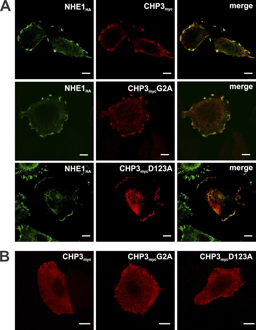
FIGURE 2.
N-Myristoylation (G2A) and Ca2+ binding (D123A)-defective mutants of CHP3 colocalize with NHE1HA at the plasma membrane. Immunofluorescence confocal microscopy of AP-1 cells stably expressing NHE1HA and transiently transfected with either wild-type or mutant forms (G2A, D123A) of CHP3myc (A) or AP-1 cells transiently transfected with the CHP3myc constructs in the absence of NHE1 (B). Subcellular distribution of NHE1HA was visualized using mouse monoclonal antibodies specific to the HA epitope followed by labeling with a goat anti-mouse secondary antibody conjugated to AlexaFluorTM-488. CHP3myc distribution was identified through a primary rabbit polyclonal antibody specific to the Myc epitope followed by a secondary goat anti-rabbit antibody conjugated to AlexaFluorTM-568. Overlapping signals in the merged images are shown in yellow. Data are representative of between two and four independent experiments. Scale bars at the bottom right of each panel represent 10 μm.

