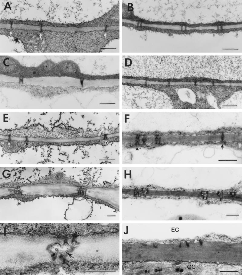
Figure 2.
Structure and development of plasmodesmata between epidermal cells during tobacco leaf maturation. A, Primary plasmodesmata (arrows) in the base of the first leaf (see Fig. 1 for numbering of leaves). B, Primary plasmodesmata in the tip of the first leaf. C, H- and Y-shaped branched plasmodesmata in the tip of the first leaf. D, Primary plasmodesmata in the base of the second leaf. E, Primary (arrows) and branched (arrowhead) plasmodesmata in the base of the second leaf. F, Primary (arrow) and branched (arrowhead) plasmodesmata in the tip of the second leaf. G, Branched plasmodesmata in the tip of the second leaf. H, Branched plasmodesmata in the base of the third leaf. I, Branched plasmodesmata in the tip of the third leaf. Note the presence of a central cavity in the plasmodesmata (arrow). J, Truncated plasmodesmata between an epidermal cell (EC) and a mature guard cell (GC). Scale bars = 0.5 μm.

