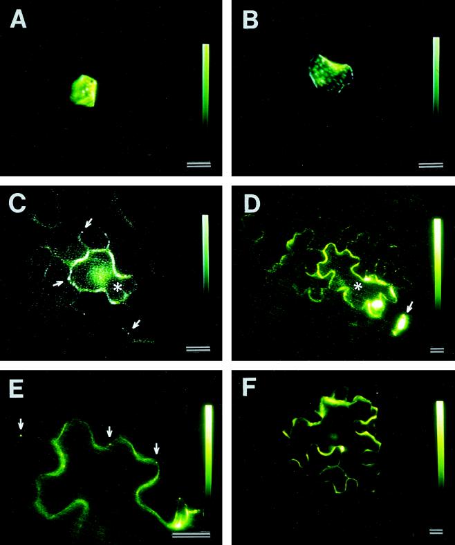
Figure 7.
Cell-to-cell trafficking of CMV 3a MP:GFP in tobacco leaf epidermis as a function of leaf development. The fusion protein was produced upon biolistic bombardment of the 3a:GFP fusion gene. A, 3a MP:GFP produced in an epidermal cell in the tip of the first leaf, which does not traffic into neighboring cells. B, 3a MP:GFP produced in an epidermal cell in the base of the second leaf, which also remains in a single cell. C, 3a MP:GFP produced in an epidermal cell (asterisk) in the tip of the second leaf, which trafficks into neighboring cells. Note the presence of green fluorescent dots in the walls of the cell producing the fusion protein and in neighboring cells (arrows). D, 3a MP:GFP produced in the base of the third leaf, which trafficks from cell to cell. The asterisk denotes the cell producing the protein. The arrow points to a guard cell that produces 3a MP:GFP but does not permit cell-to-cell trafficking of the fusion protein. E, High-magnification view of D showing the presence of fluorescent dots in the cell walls. F, 3a MP:GFP produced in an epidermal cell in the base of the third leaf. The fusion protein was not targeted to complex secondary plasmodesmata, as determined by the lack of green fluorescent dots in the cell walls. The protein does not traffic into neighboring cells. Scale bars = 20 μm.

