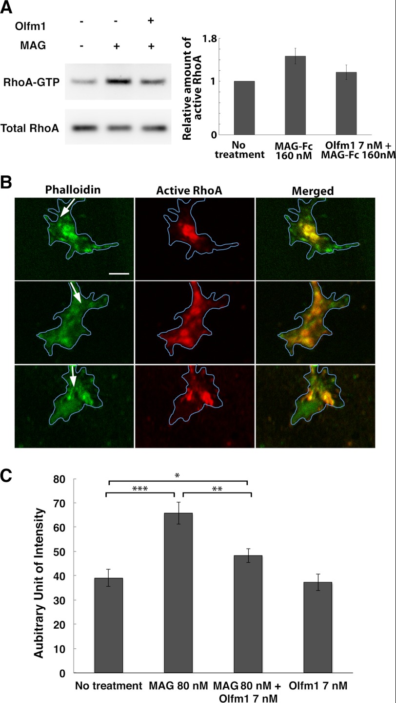
FIGURE 7.
Inhibition of NgR1-mediated and MAG-induced RhoA activation by Olfm1. A, COS7 cells were transfected with cDNAs encoding NgR1, p75 NTR and/or LINGO-1. 48 h after the transfection, purified Olfm1 or buffer control was added for 10 min, and then cells were stimulated with MAG-Fc (160 nm). Cells were harvested 15 min later, and the active form of RhoA (RhoA-GTP) was pulled down and detected by Western blotting. These experiments were repeated seven times. B, DRG growth cones were treated with MAG (80 nm) for 10 min with (lower panels) or without (center panels) Olfm1 (7 nm) pretreatment. Upper panels, untreated control. The explants were stained with anti-active RhoA antibody (red) together with phalloidin (green). Arrows show the direction of axon growth. Blue lines represent the outlines of growth cones. Scale bar = 5 μm. C, quantification of the fluorescence intensities for active RhoA in the immunostained growth cones (>30 growth cones from three to six explants) measured using ImageJ. *, p < 0.05; **, p < 0.01; ***, p < 0.001.

