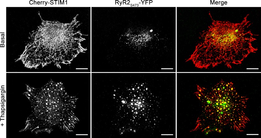
FIGURE 5.
RyR co-localize with STIM1 puncta following store depletion in HEK293 cells. Confocal images of HEK293 cells co-overexpressing Cherry-STIM1 and RyR2S437-YFP recorded in red and green channels, respectively, prior to store depletion (top panels) and 10 min following incubation in bath solution supplemented with 1 μm thapsigargin (bottom panels). Right columns show merged images of Cherry-STIM1 and RyR2S437-YFP. Co-localization is depicted in yellow. Scale bar, 10 μm. Images were taken at the bottom of the cells; optical slice depth was <2.5 μm. Results shown are representative images from eight independent experiments.

