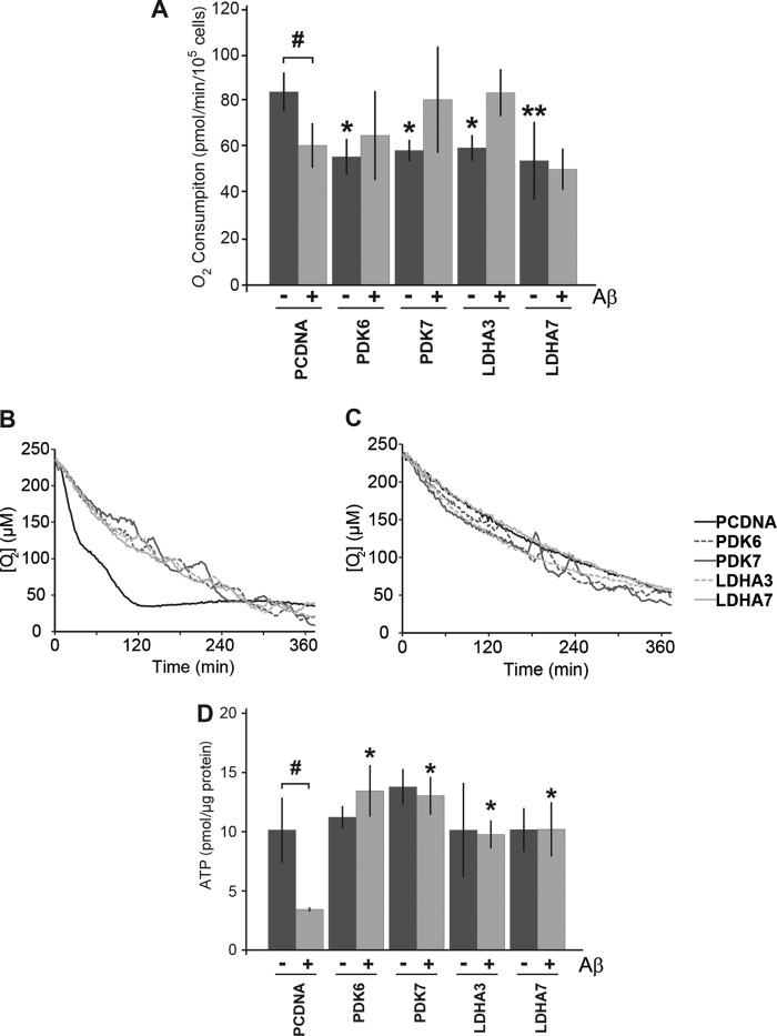
FIGURE 3.
Respiration is decreased but ATP levels are maintained in cells overexpressing LDHA and PDK1. A, oxygen consumption was monitored in B12 cell lines using the MitoXpress-Xtra-HS fluorescent probe. All clonal cell lines overexpressing LDHA and PDK1 displayed significantly lower levels of oxygen consumption under control conditions compared with cells expressing an empty vector (pcDNA) (*, p < 0.05; **, p < 0.01). Oxygen consumption significantly decreased in pcDNA control cells following 48 h of treatment with Aβ(25–35) (20 μm) compared with untreated conditions (#, p < 0.05). In contrast, cells overexpressing LDHA or PDK1 maintain or increase their oxygen consumption following 48 h of Aβ exposure. B, representative example of oxygen consumption over time for the indicated B12 cell lines. C, representative example of oxygen consumption over time following 48 h of Aβ(25–35) (20 μm) treatment. D, cells overexpressing LDHA or PDK1 had similar levels of ATP when compared with control cells under normal culture conditions. Cells expressing empty vector had significantly lower levels of ATP following exposure to Aβ (#, p < 0.05), whereas LDHA- and PDK1-overexpressing cells maintained significantly higher ATP levels than the control following treatment with Aβ (*, p < 0.05). Data represent the average ± S.D. of three independent experiments. Data were analyzed by a one-way ANOVA followed by a Tukey test.

