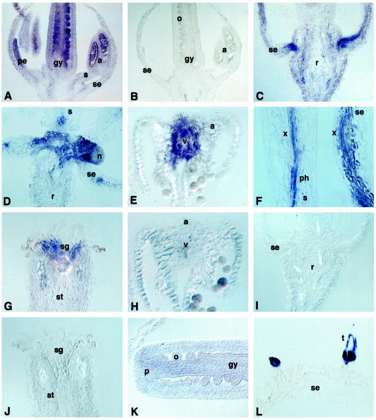
Figure 2.
Localization of MT1a and MT2a in Arabidopsis flowers. A and C to G, Sections hybridized with MT1a antisense riboprobe; B, section probed with MT1a sense; H to L, sections probed with MT2a antisense. A, B, and K, Flowers at stage 8 to 9. C to J and L, Flowers at anthesis. All sections are from plants grown without excess copper. a, Anthers; gy, gynoecium; n, nectary; o, ovule; p, placenta; pe, petal; ph, phloem; r, receptacle; s, stamen; se, sepal; sg, stigma; st, style; t, trichome; v; vascular bundle; x, xylem.

