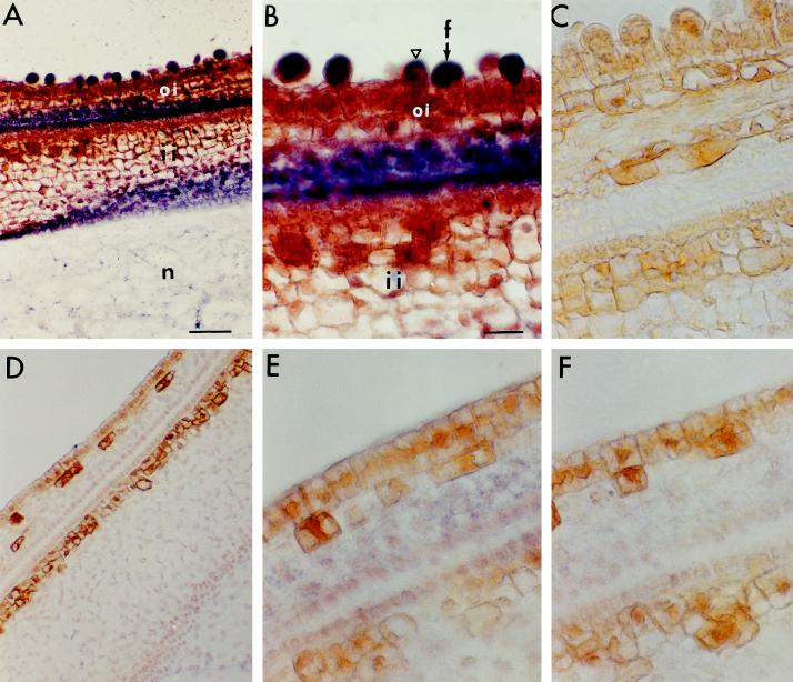
Figure 2.
In situ hybridization of SuSy in cross-sections of FLS (A–C) and fls (D–F) ovules. The purple signals represent SuSy mRNA. A, B, D, and E, Cross-sections were hybridized with an antisense RNA probe generated from SS3 cDNA. Note in B the very strong SuSy mRNA signals in the large and spherically shaped initiating fiber cells (arrow), the weak signals in the small fiber cells (triangle), and the undetectable signals in the nondifferentiating epidermal cells. Also note in E that SuSy mRNA was undetectable in the nondifferentiating epidermis of the fls mutant. C and F, Cross-sections were hybridized with sense RNA probe. Bars in A and B are 50 and 22 μm, respectively (magnifications of A and D, B and C, and E and F). f, Fiber cell; oi, outer integument; ii, inner integument; n, nucellus.

