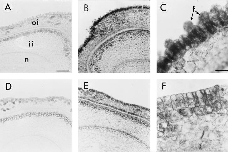
Figure 3.
Immunogold localization of SuSy protein in cross-sections of FLS (A–C) and fls (D–F) ovules. A and D, Cross-sections were treated with preimmune serum. B, C, E, and F, Cross-sections were treated with polyclonal antibody against SuSy. Note in C the very strong SuSy protein signals in initiating fiber cells (arrows) and weak signals in the nondifferentiating epidermal cells (see the cell between the two initiating fiber cells indicated by arrows). Also note in F that very little SuSy protein can be detected in the ovule epidermis of the fls mutant. Bars in A and C are 77 and 19 μm, respectively (magnifications of A and B, D and E, and C and F). f, Fiber cells; oi, outer integument; ii, inner integument; n, nucellus.

