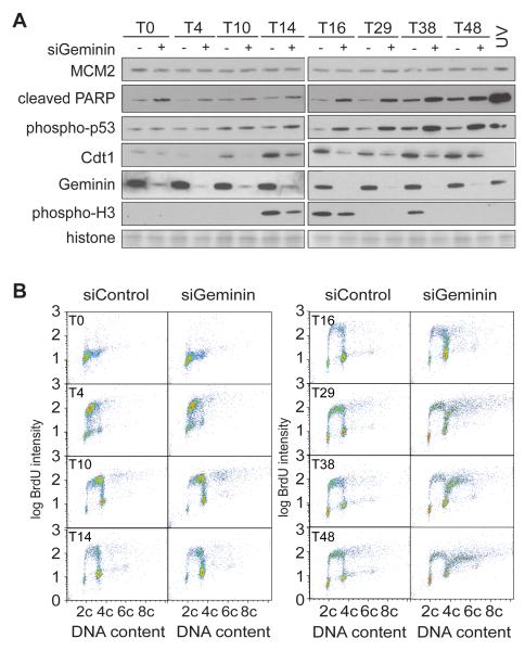
Figure 2. Flow cytometry of cells re-replicating after geminin depletion.
Cells were released from a double thymidine block after prior treatment with either control or geminin RNAi, as in Figure 1A. A. Western blot analysis of whole cell extracts at different times after thymidine release. Geminin RNAi: +; control RNAi: −. As control, cells were treated with 120 mJ UV 4h after thymidine release. The membrane was also stained with amido black to show equal loading of histones. B. At different times after release from double thymidine block, cells were pulsed with BrdU and analysed by flow cytometry.

