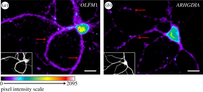
Figure 2.
In situ hybridization reveals different patterns of localization in neuronal dendrites. Fluorescent microscopic evaluation of biotin-conjugated oligoprobes on paraformaldehyde-fixed 14-day-cultured mouse cortical neurons hybridized with biotin-conjugated 25mer-oligoprobes detected with streptavidin-Alexa568. For each image, the small bottom left corner panels represent MAP2 immuno-staining. Patterns of distribution are highlighted with red arrows. (a) Probe against OLFM1 transcript illustrates a uniform distribution in dendrites; (b) probe against ARHGDIA transcript illustrates a punctated distribution in dendrites.

