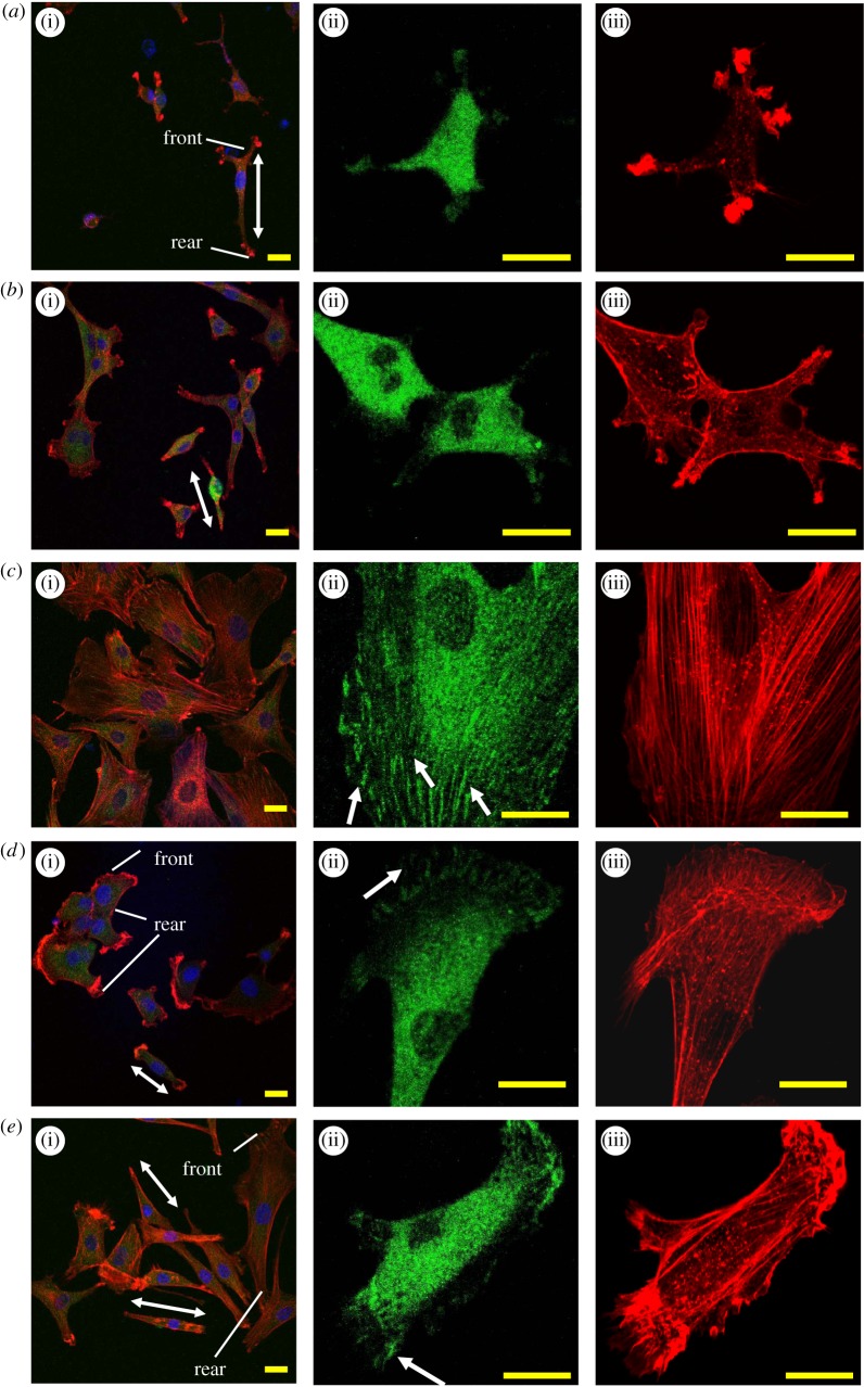
Figure 3.
Morphology and focal contact formation of the SMCs cultured on the salt-treated multilayers for 24 h. Representative CLSM images of SMCs on the multilayers treated with (a) 1, (b) 2, (c) 3, (d) 4 and (e) 5 M NaCl solutions, respectively. (a(i)–e(i)) Merge fluorescence images of vinculin (green), actin (red) and nucleus (blue). (a(ii)–e(ii)) and (a(iii)–e(iii)) vinculin and actin in a single cell, respectively. Arrows in (c(ii)), (d(ii)) and (e(ii)) indicate areas of large focal adhesion plaques. Two-head arrows indicate the polarity of the SMCs, and the texts illustrate the cell front (cell leading edge) and rear.

