Figure 2. .
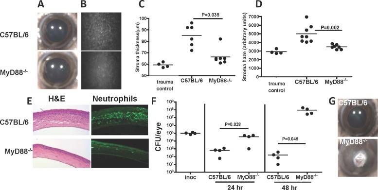
The role of MyD88 in S. marcescens–induced corneal infection. Corneas of C57BL/6 and MyD88−/− mice were abraded, and 1 × 10⁷ S. marcescens clinical isolate (056) in 2 μL PBS were added topically. A 2 mm diameter punch from a silicone hydrogel contact lens then was placed on the corneal surface for 2 hours. Corneal images were taken by light microscopy, and corneal thickness and haze (B–E) were examined using the ConfoScan3 microscope system. (A) Representative corneas 24 hours after infection (original magnification is ×20). (B) Representative Confoscan images of the central corneal stroma (original magnification is ×200). (C, D) Corneal thickness and haze measured by Confoscan. Trauma controls were abraded and exposed to sterile PBS. (E) Representative 5 μm corneal sections stained with hematoxylin and eosin or immunostained with the anti-mouse neutrophil Ab NIMP R14. (F) CFU at 24 and 48 hours post infection. Inoc: CFU after 2 hours on the ocular surface, representing the inoculum). (G) Representative C57BL/6 and MyD88−/− corneas 48 hours post infection. Data points represent individual corneas; the experiment was repeated twice with similar results.

