Fig. 2.
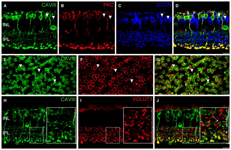
CAVIII is localized to rod bipolar cells (RBCs) and a subset of cone bipolar cells in the wild-type mouse retina. (A–D) Triple-labeling for CAVIII, the RBC marker, PKC and the cone bipolar cell marker, SCGN in a vertical section of the mouse retina. Merged image shown in (D). Most CAVIII-positive bipolar cells colocalize with PKC; however, those lacking PKC colocalize with SCGN, indicating that they are cone bipolar cells (arrowheads). (E–G) Staining of a mouse retinal whole-mount labeled for CAVIII and PKC, focused at the level of the proximal INL. Merged image shown in (G). As in vertical sections, most CAVIII-positive bipolar cells are PKC positive, whilst a smaller number are PKC negative (arrowheads). (H–J) Vertical section labeled for CAVIII and vGluT1. The inset shows a magnified view of the region defined by the dotted rectangle. The inset in (C) shows the five strata (1–5) of the IPL. CAVIII colocalizes with vGluT1 in the middle region of the IPL (S3), as well as in S5 where the RBCs stratify (arrows). Scale bars: 10 μm. CAVIII, carbonic anhydrase-related protein VIII; INL, inner nuclear layer; IPL, inner plexiform layer; PKC, protein kinase C; SCGN, secretagogin; vGluT1, vesicular glutamate transporter 1.

