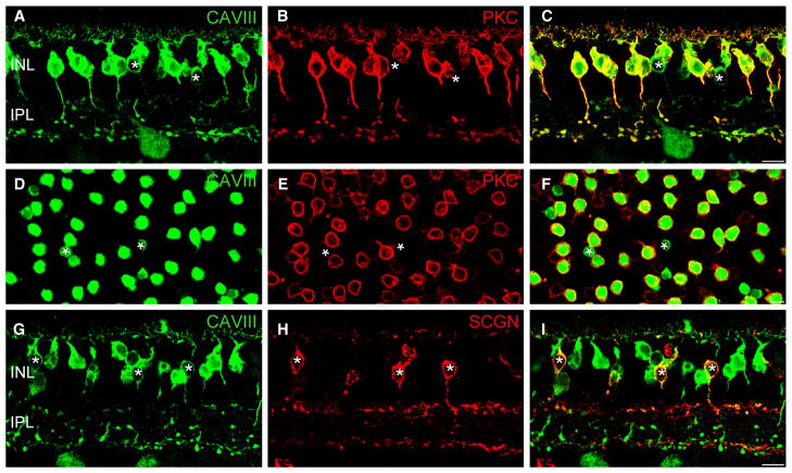
Fig. 4.
CAVIII is localized to RBCs and a subset of cone bipolar cells in the macaque retina. (A–F) Immunolocalization of CAVIII and PKC in vertical section (AC) and a whole-mount (D–F) of the macaque retina. The whole-mount image is taken with the focus at the levels of the distal INL. As in the mouse retina, most CAVIII-positive bipolar cells are also immunoreactive for PKC, indicating that they are RBCs (C and F). Asterisks indicate CAVIII-positive bipolar cells that lack PKC staining. (G–I) Vertical section labeled for CAVIII and SCGN shows CAVIII-positive bipolar cells that stratify in the outermost region of the IPL, indicating that CAVIII is present in DB1 OFF cone bipolar cells (asterisks). Scale bars: 10 μm. CAVIII, carbonic anhydrase-related protein VIII; INL, inner nuclear layer; IPL, inner plexiform layer; PKC, protein kinase C; SCGN, secretagogin.

